Autologous Micrografting and Fibroblasts in Regenerative Medicine
Autologous Micrografting is the procedure of taking a small skin graft and using that tissue to stimulate the growth and creation of healthy cells on the derma – this allows chronic wounds to heal, helps reduce the visibility of scars, treats alopecia or hair loss, or enables improved recovery from dentistry surgery.
The Skin Structure
Autologous Micrografting is the procedure of taking a small skin graft and using that tissue to stimulate the growth and creation of healthy cells on the derma – this allows chronic wounds to heal, helps reduce the visibility of scars, treats alopecia or hair loss, or enables improved recovery from dentistry surgery.
The skin is a complex organ composed of various cells and glands, each performing distinct functions. It is intricately held together by proteins and nourished with oxygen and nutrients. Because of this complexity, merely transferring skin tissue from one part of the body to another does not naturally support effective regenerative processes.
Successful skin regeneration requires more than just tissue placement; it involves a comprehensive understanding of the skin's intricate structure and the biological processes that sustain its function.
Why is Micrografting Useful?
When harvesting a micrograft, we collect a tissue sample rich in diverse cell types. Key components of this graft include:
- Dermal Fibroblasts: These cells are crucial for the structural integrity and repair of the skin. They produce collagen and other extracellular matrix proteins that support skin regeneration.
- Mesenchymal Stem Cells (MSCs): These progenitor cells have a vital role in skin regeneration. They can differentiate into various cell types and replace damaged cells or transform into cells needed in the affected area.
In essence, the inclusion of dermal fibroblasts and mesenchymal stem cells in micrografts enhances their regenerative potential, making them effective for repairing and rejuvenating the skin.
You can find out more about stem cells, by clicking the link here.
Dermal Fibroblasts perform another uniquely important role; they are responsible for building structures in our tissue and are hugely important for wound healing. By making and releasing collagen fibres, elastic fibres, and sugar-based glycosaminoglycans, other cells (such as epithelials) can be easily bound and held together in a wider scaffold. The impact of dermal fibroblasts and mesenchymal stem cells cannot be overstated. Without their presence, new tissue formed after an injury would struggle to maintain its structure, receive essential nutrients, and prevent cell death (necrosis).
In addition to this, fibroblasts are known to play a critical role in the immune response to tissue damage. By initiating inflammation responses when confronted with foreign microorganisms their response also starts a cascade of functions which eliminates these foreign threats. Without this cascade, the body will not replenish dead or dying cells and will create a perfect environment for the skin to heal. On the opposite side, fibroblasts will also regulate the body in the opposite way and suppress the immune response in tumours.
How to Conduct a T-Lab Micrografting Treatment
T-Lab has developed a ground-breaking technique for breaking down tissues into their fundamental components while preserving cell viability. This method results in an injectable solution enriched with viable mesenchymal stem cells and fibroblasts, leading to significant clinical benefits.
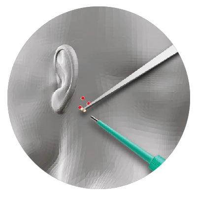
Graft Collection: The process begins with harvesting a skin graft rich in both fibroblasts and mesenchymal stem cells. Optimal sites for collection include:
This approach ensures that the extracted cells are maximally effective for therapeutic applications, promoting enhanced healing and regeneration.
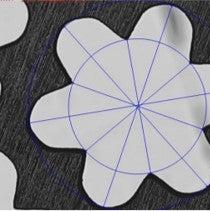
After collecting the skin grafts, the next crucial step is to separate viable fibroblasts and stem cells from the surrounding tissue—a process that has traditionally been quite challenging. T-Lab has overcome this hurdle with their advanced technology known as the Microlyser.
How the Microlyser Works:
- Technology: The Microlyser utilizes hundreds of honeycomb-shaped blades to cut the tissue and induce shear stress simultaneously. This process efficiently isolates the fibroblasts and stem cells.
- Blade Specifications: The blades used in the Microlyser are microscopic, with sizes ranging from 40 microns to 3000 microns, tailored to meet specific requirements.
When a skin graft is processed through a series of Microlysers, the technology efficiently breaks down the cells, allowing them to float freely in a PRP or saline mixture. At the same time, the Microlysers act as a filter, removing larger, unwanted cells to ensure that only the beneficial components are injected into the patient.
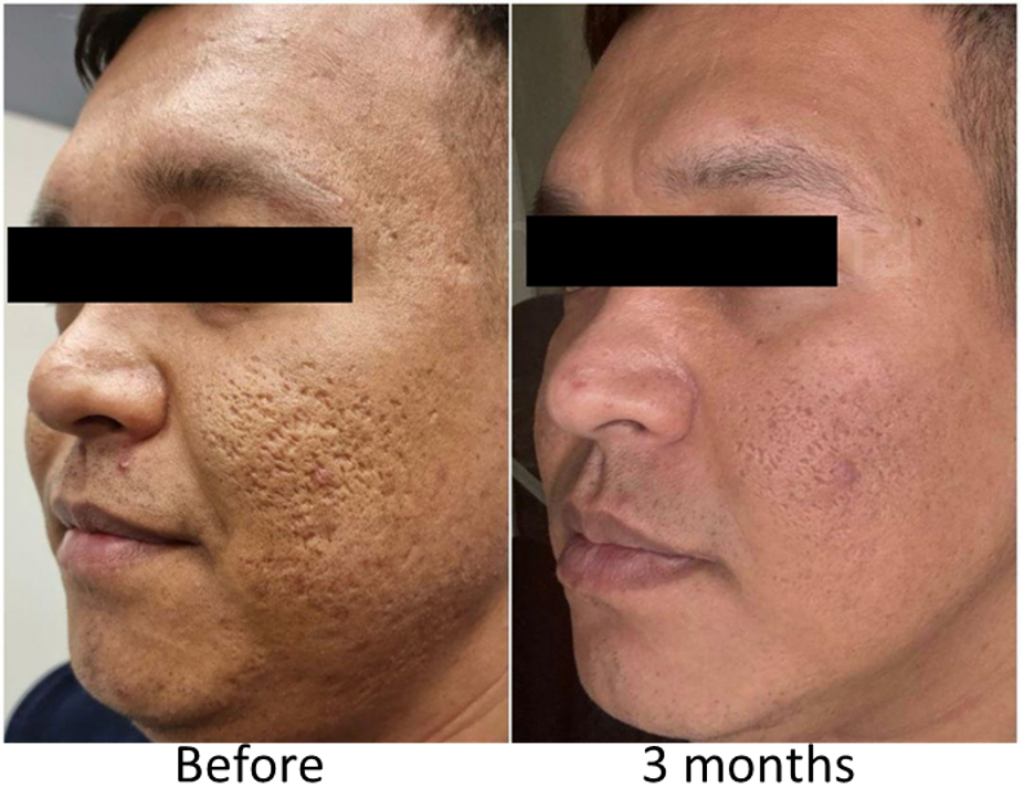
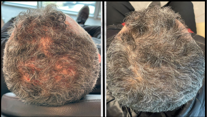
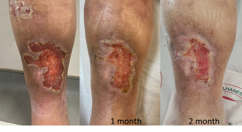
The results of this process are impressive and are reflected in various clinical applications. Below are images showcasing the benefits of using the Microlyser technology for different treatments:
